FIG 1.
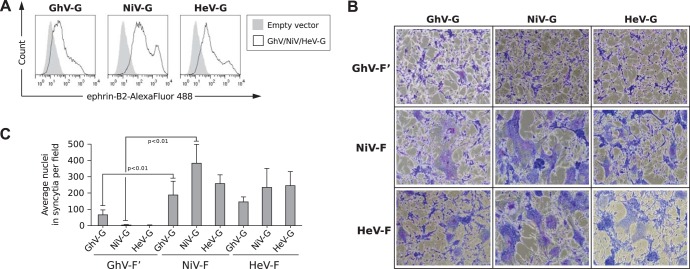
Functional properties of GhV envelope glycoproteins. (A) HNV-G proteins bind ephrin-B2. HEK293T cells were transfected with GhV-G, NiV-G, or HeV-G expression plasmids. Empty pcDNA3.1− vector was also transfected to serve as a negative control. At 27 h posttransfection, cells were processed for fluorescence-activated cell sorting (FACS) analysis using soluble ephrin-B2-(hu)Fc as the primary staining agent and Alexa Fluor 488-conjugated goat anti-human IgG (Fc) as the secondary antibody. (B) Fusogenicity of GhV-F′ and GhV-G proteins. Human U87 glioblastoma cells were transfected with plasmids encoding the indicated F (GhV-F′, NiV-F, and HeV-F) and G (GhV-G, NiV-G, and HeV-G) proteins at a 1:1 ratio, such that all homotypic and heterotypic combinations are represented. Cells were fixed and stained with Giemsa at 30 h posttransfection. Images were taken from 5 random fields for each condition. Representative images are shown. (C) Quantification of fusogenicity of GhV-F′ and GhV-G proteins. The images were quantified by manually counting the nuclei in syncytia in each field using ImageJ counting software. Results are presented as means and standard deviations (SD) from 5 fields. Significant differences were observed between GhV-F′ and NiV-F (P < 0.01; one-tailed Student t test).

