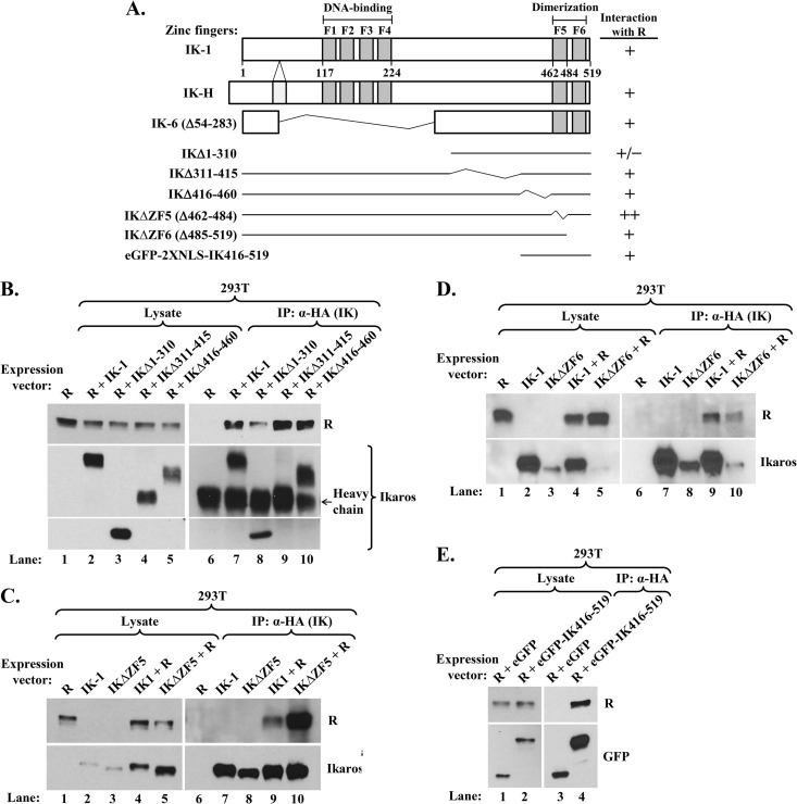
FIG 8.
Ikaros domains involved in its interaction with R. (A) Schematic diagrams showing structures of IK-1, IK-H, IK-6, and deletion variants studied here. Numbers indicate amino acid residues. F1 to F6 denote zinc fingers. +/−, +, and ++ denote interaction with R that was less than, similar to, or greater than that observed with IK-1, respectively. (B, C, and D) Immunoblots showing coimmunoprecipitation of R with Ikaros deletion variants. (B) 293T cells in 6-well plates were cotransfected as follows: lanes 1 and 6, 0.1 μg pcDNA3-R; lanes 2 and 7, 0.1 μg pcDNA3-R plus 0.2 μg pcDNA3-HA-IK-1; lanes 3 and 8, 0.1 μg pcDNA3-R plus 0.9 μg pcDNA3-HA-IKΔ1-310; lanes 4 and 9, 0.1 μg pcDNA3-R plus 0.9 μg pcDNA3-HA-IKΔ311-415; and lanes 5 and 10, 0.1 μg pcDNA3-R plus 0.9 μg pcDNA3-HA-IKΔ416-460; total DNA was brought up to 1.0 μg per well with pcDNA3.1 where needed. Whole-cell extracts were prepared 48 h later, and protein was immunoprecipitated with anti-HA tag antibody. (C) 293T cells in 6-well plates were cotransfected as follows: lanes 1 and 6, 0.2 μg pcDNA3-R; lanes 2 and 7, 0.2 μg pcDNA3-HA-IK-1; lanes 3 and 8, 0.2 μg pcDNA3-HA-IKΔZF5; lanes 4 and 9, 0.2 μg pcDNA3-R plus 0.36 μg pcDNA3-HA-IK-1; and lanes 5 and 10, 0.2 μg pcDNA3-R plus 0.36 μg pcDNA3-HA-IKΔZF5; total DNA was brought up to 0.56 μg per well with pcDNA3.1 where needed. Whole-cell extracts were processed as described above. (D) 293T cells were cotransfected and processed as described for the experiment whose results are shown in panel C, except with pcDNA3-HA-IKΔZF6 in place of pcDNA3-HA-IKΔZF5. (E) Immunoblot showing coimmunoprecipitation of R with eGFP-fused IK416-519. 293T cells in 6-well plates were cotransfected with 0.1 μg pcDNA3-R and 0.9 μg pcDNA3-HA-eGFP-2XNLS or 0.2 μg pcDNA3-HA-eGFP-2XNLS-IK416-519 plus 0.7 μg pcDNA3.1. Whole-cell extracts were processed as described above, except that blots were probed with anti-GFP antibody.

