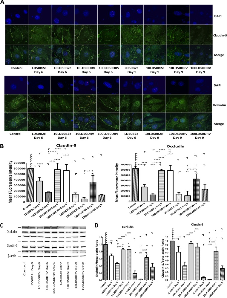
FIG 4.
Effects of brain extracts derived from mice infected with CVS-B2c or DRV-Mexico on the expression of TJ proteins in BMECs. (A) Mouse BMECs were cocultured with brain extracts from mice infected with different doses of CVS-B2c or DRV-Mexico. After 24 h, BMECs were fixed and were stained with DAPI and an anti-claudin-5 antibody or with DAPI and an anti-occludin antibody. The staining was visualized by confocal microscopy. (B) MFI for the ROI drawn around the cells and statistical analysis. (C) The expression of TJ proteins in BMECs after coculturing with brain extracts was also detected using Western blotting. (D) TJ protein expression as detected by WB was quantitatively analyzed in BMECs treated with brain extracts from mice infected with either virus (CVS-B2c or DRV-Mexico). bEnd.3 cells treated with brain extracts from sham-infected mice were included as controls. Data are means ± SEM of results from three independent experiments. Statistical analyses in panels B and D were performed with one-way ANOVA followed by Tukey's post hoc test. Asterisks indicate statistical significance (*, P < 0.05; **, P < 0.01; ***, P < 0.001; ****, P < 0.0001). Three samples were collected from three replicates in each group for statistical analysis.

