FIG 2.
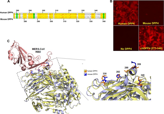
Blades IV and V from the β-propeller of hDPP4 make mDPP4 permissible to MERS-CoV infection. (A) Vector NTI protein sequence alignment of human (top strand) and mouse (bottom strand) DPP4 molecules. Yellow highlighted regions indicate conserved amino acids, white regions signify amino acids that are functionally different (i.e., hydrophobic and hydrophilic), and green highlighting indicates amino acids that are different but functionally similar (i.e., the threonine and serine are both polar and uncharged). (B) HEK 293T cells were transfected with the indicated DPP4. At ∼20 h posttransfection, cells were infected with rMERS-CoV-red virus at an MOI of 5, and infection was assessed ∼18 h postinfection by fluorescence microscopy. (C) 3D molecular PyMOL software was employed to visualize the mDPP4 structure overlaid onto the hDPP4 structure. The hDPP4 structure was based upon the crystal structure resolved in context with the MERS S RBD (PDB code 4L72). MERS S protein is displayed in red, hDPP4 in yellow, and mDPP4 in blue. The mDPP4 sequence was threaded using the I-TASSER software (11). The expanded view depicts the DPP4 region at the interaction surface. Numbered and highlighted are the specific amino acids chosen for mutation in the mDPP4 protein.

