
FIG 4.
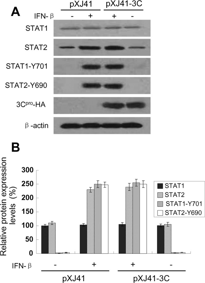
Protein level and phosphorylation status of STAT1 and STAT2 in FMDV 3Cpro-expressing cells after IFN-β stimulation. (A) HeLa cells were transfected with empty vector pXJ41 or pXJ41-3C. At 24 h posttransfection, cells were either left untreated (−) or treated (+) with 1,000 U/ml of IFN-β for 1 h and harvested for Western blotting with antibody against STAT1, STAT2, phospho-STAT1 (STAT1-Y701), phospho-STAT2 (STAT2-Y690) or HA, as indicated. The same blot was incubated with β-actin antibody as a protein loading control. The data presented here are results from one experiment of three Western blotting experiments. (B) Densitometry analysis of the digital image from three independent experiments. The band intensities are shown as the relative protein expression levels, normalized with β-actin. Error bars indicate the standard deviations of three experiments.