
FIG 7.
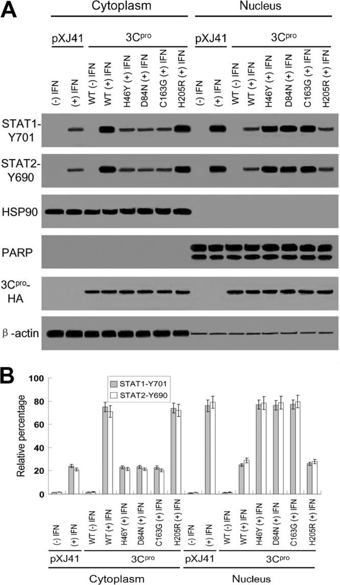
(A) Phosphorylated STAT1 and STAT2 in nuclear and cytoplasmic fractions. Subcellular fractionation of HeLa cells, 1 h after IFN-β treatment, was performed for nuclear and cytoplasmic fractions, followed by Western blotting using STAT1-Y701 antibody, STAT2-Y690 antibody, anti-HSP90 antibody as a cytoplasmic marker, and anti-PARP antibody as a nuclear protein marker, as indicated. HA antibody was used to detect FMDV 3Cpro expression (3Cpro-HA), and β-actin antibody was used as a loading control. (B) Densitometry analysis of the digital image of phosphorylated STAT1 and STAT2 in nuclear and cytoplasmic fractions from three independent experiments. The band intensities of each fraction are shown as the relative percentages of the total density of corresponding cytoplasmic and nuclear fractions. Normalization for cytoplasmic and nuclear fractions was done with HSP90 and PARP, respectively. Error bars indicate the standard deviations of three experiments. −, without IFN-β treatment; +, with IFN-β treatment.