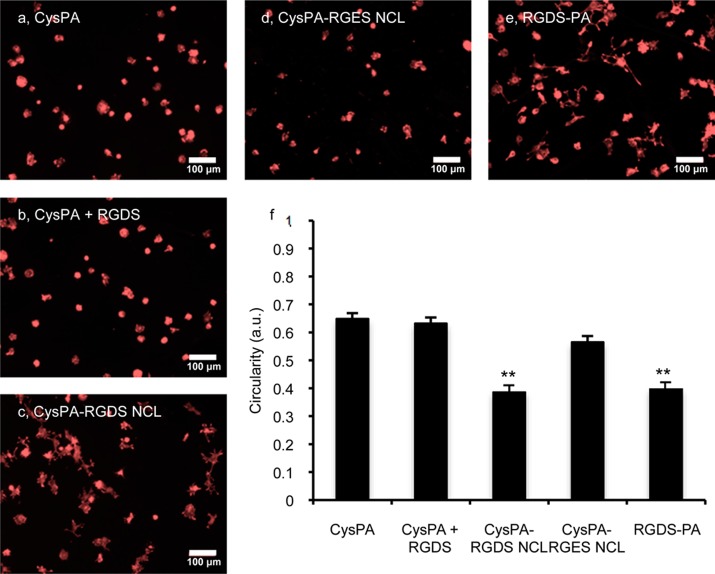
Figure 4.
Fibroblast morphology on PA-coated coverslips. (a–e) Fluorescent micrographs of NIH/3T3 fibroblasts cultured for 5 h and stained by phalloidin for F-actin on coverslips coated with CysPA (a), CysPA and soluble RGDS peptide (b), NCL RGDS-PA (c), NCL RGES-PA (d), and presynthesized RGDS-PA not formed by NCL (e). (f) Comparison of cell circularity (4π × area/perimeter2) on various PA-coated surfaces (** p < 0.01 vs CysPA, n = 100 cells).

