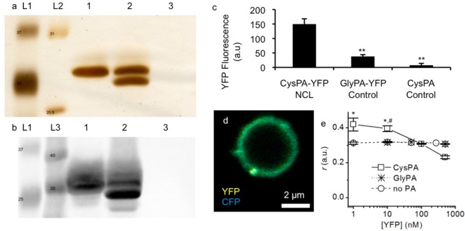
Figure 6.
Characterization of PA–protein conjugates formed by NCL. (a,b) SDS-PAGE visualized by silver staining (a) and Western blot against GFP (b) were used to analyze the NCL reaction between YFP and PA (Lane 1: YFP only, Lane 2: YFP conjugated to CysPA, Lane 3: CysPA only, Lanes L1, L2, L3: protein standards with indicated molecular weights in kDa). (c) YFP fluorescence intensity after conjugation to CysPA-coated surface (CysPA-YFP NCL). An identical PA lacking cysteine (GlyPA-YFP Control) or CysPA treated with reaction buffer alone (CysPA Control) was used as a control (** p < 0.01 vs CysPA-YFP NCL). (d) Confocal fluorescence microscopy of CysPA nanofiber-coated alginate microparticles simultaneously reacted with YFP and CFP. (e) Fluorescence anisotropy of YFP following NCL reaction with CysPA. GlyPA was used as a control (* p < 0.05 vs YFP; #p < 0.05 vs GlyPA).

