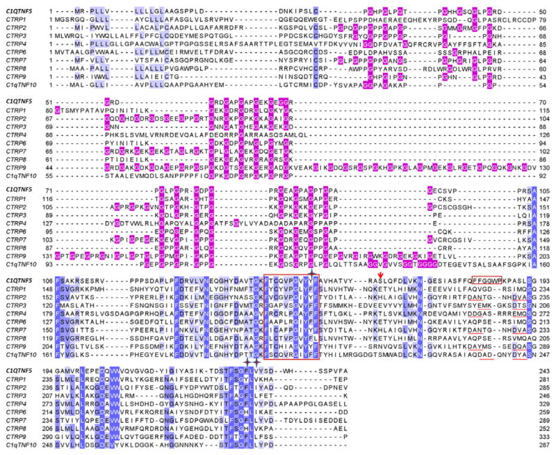
Fig. 5.
Sequence comparisons of human C1QTNF5 with its paralogues. Conserved residues of the gC1q domain are highlighted with white or black letters on a blue background according to their conservation. Gly residues in the collagen domain are highlighted with white font on a red background. The conserved hydrophobic motif (residues 143–153) and the characteristic hydrophobic segment FFGGWP (residues 181–186) of human C1QTNF5 are outlined with red rectangles. Residues forming the hydrophobic core at the trimerization interface are indicated with stars. The pathogenic mutation site of C1QTNF5 is indicated with a red arrow. Structural elements coordinating with Ca2+ ions are highlighted with red underlining.

