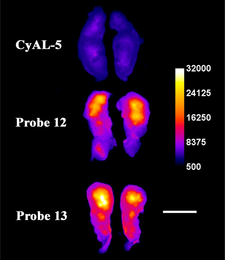
Figure 7.

Representative ex vivo, deep-red fluorescence images of excised and sliced rat prostate tumors from animals sacrificed at 24 h after intravenous dosing with deep-red CyAL-5 dye, probe 12, or probe 13 (3.0 mg/kg). Each tumor is sliced along the longest axis with the core of the tumor facing the camera. The fluorescence intensity scale bar applies to all images (arbitrary units). Length scale bar = 1 cm.
