Figure 1.
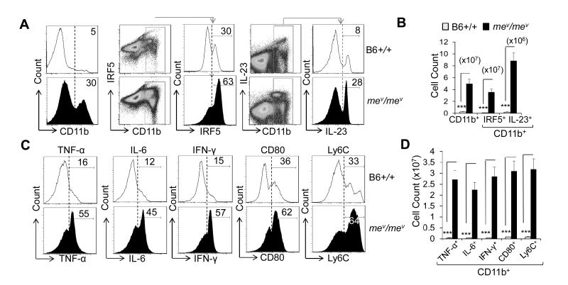
Increased inflammatory macrophage counts in B6-mev/mev mice. A, Percentage of CD11b+ (left), CD11b+ interferon regulatory factor 5–positive (IRF-5+) (middle), and CD11b+ interleukin-23–positive (IL-23+) (right) macrophages in a single-cell suspension of spleen from B6-+/+ and B6-mev/mev mice as determined by fluorescence-activated cell sorting (FACS) analysis. B, Cell counts of the indicated populations in the spleens of B6-+/+ and B6-mev/mev mice. C, Percentage of intracellular tumor necrosis factor α (TNF-α), IL-6, interferon-γ (IFN-γ), and surface CD80 and lymphocyte antigen (Ly6C) expression on the CD11b+ subpopulation of spleen cells, as determined by FACS analysis. D, Total cell count of CD11b+ macrophages that express the indicated intracellular or surface molecules, as determined by multiplying the total single-cell suspension count by the percentage in the gated population. Values are the mean (± SEM) (n = 5 mice per group). *** = P < 0.001.

