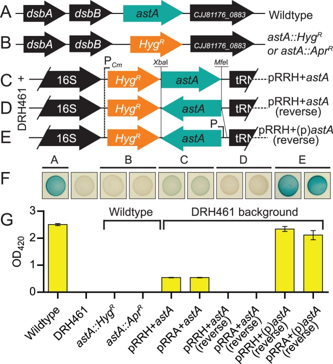
Figure 3. Mutagenesis of the arylsulfatase gene astA with aph(7″) or aac(3)IV non-polar markers and complementation of ΔastA via genomic insertion with pRRH or pRRA.
(A) Loci arrangement of astA single-gene operon in C. jejuni 81–176. (B) Deletion of astA with either aph(7″) or aac(3)IV from pAC1H or pAC1H. (C) Introduction of promoterless astA into pRRH or pRRA in the same orientation as the cat promoter created pRRH+astA or pRRA+astA and resulted in polycistronic expression of astA with aph(7″) or aac(3)IV. (D) Promoterless astA inserted in the opposite orientation to the cat promoter (designated pRRH+astA (reverse) or pRRA+astA (reverse) (E) Insertion of the endogenous astA promoter and astA in the opposite orientation to the cat promoter in pRRH and pRRA created pRRH+(p)astA (reverse) and pRRA+(p)astA (reverse). Only HygR plasmids/strains are depicted in B–E, but both HygR and AprR plasmids represented with HygR in C, D and E were integrated into the genome of the ΔastA strain, DRH461. (F) Arylsulfatase activity of the deletion and complementation strains was assessed by spotting 10 µL of OD-standardized cultures onto MH agar plates supplemented with the chromogen XS cleaved by arylsulfatase. A blue-green color indicates activity, and the spots correspond to labels on the bar graph below. (G) Quantification of arylsulfatase activity from broth cultures to assess transcription of astA.

