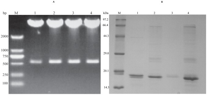
Figure 4. Double digestion and purification of VHH antibodies.
(A) Agarose gel electrophoresis of VHH antibodies double digested with BamHI and HindIII. Upper bands of 4500 bp correspond to vector pCANTAB5E whereas lower bands of 400 bp correspond to VHH fragments of P2 (lane 1), P9 (lane 2), P10 (lane 3), P13 (lane 4), respectively, and M stands for marker. (B) 12% polyacrylamide SDS electrophoresis gel of VHH antibodies purified from E. coli BL21 using a Ni-NTA column. A single band of about 15 kDa is observed for each purified VHH: P2 (lane 1), P10 (lane 2), P9 (lane 3), P13 (lane 4); M stands for marker.

