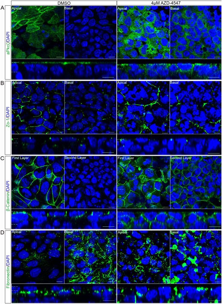
Figure 8. Inhibition of the Fgfr disrupted the normal localisation of polarity and junction proteins in the outer layer of embryoid bodies.
Embryoid bodies were treated with the Fgfr inhibitor 4 µM AZD-4547, or 0.04% DMSO for 7 days. Whole-mount immunostaining demonstrated the localisation of proteins normally polarised in the primitive endoderm. (A) aPkcζ/λ a member of a polarity complex, (B) Zo-1 a tight junction protein, (C) β-catenin a protein in the adherens junction and (D) the basement membrane protein Fibronectin, were shown to lose their apico-basolateral polarised localisation when embryoid bodies were grown with an Fgfr inhibitor suggesting a disruption in the apico-basolateral polarity of these cells. A representative image from 3 independent experiments is shown. Scale bars 10 µm. Dotted lines represent position that the relevant orthogonal or aerial images were taken.

