
Figure 1. Radiographically identifiable (A) necrotic, (B) solid, and (C) cystic node metastasis defined by axial contrast-enhanced CT scans.
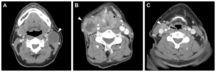
Note a contrast-enhancing thin wall and homogeneous low-density content in the cystic node metastasis.
Official websites use .gov
A
.gov website belongs to an official
government organization in the United States.
Secure .gov websites use HTTPS
A lock (
) or https:// means you've safely
connected to the .gov website. Share sensitive
information only on official, secure websites.
Note a contrast-enhancing thin wall and homogeneous low-density content in the cystic node metastasis.