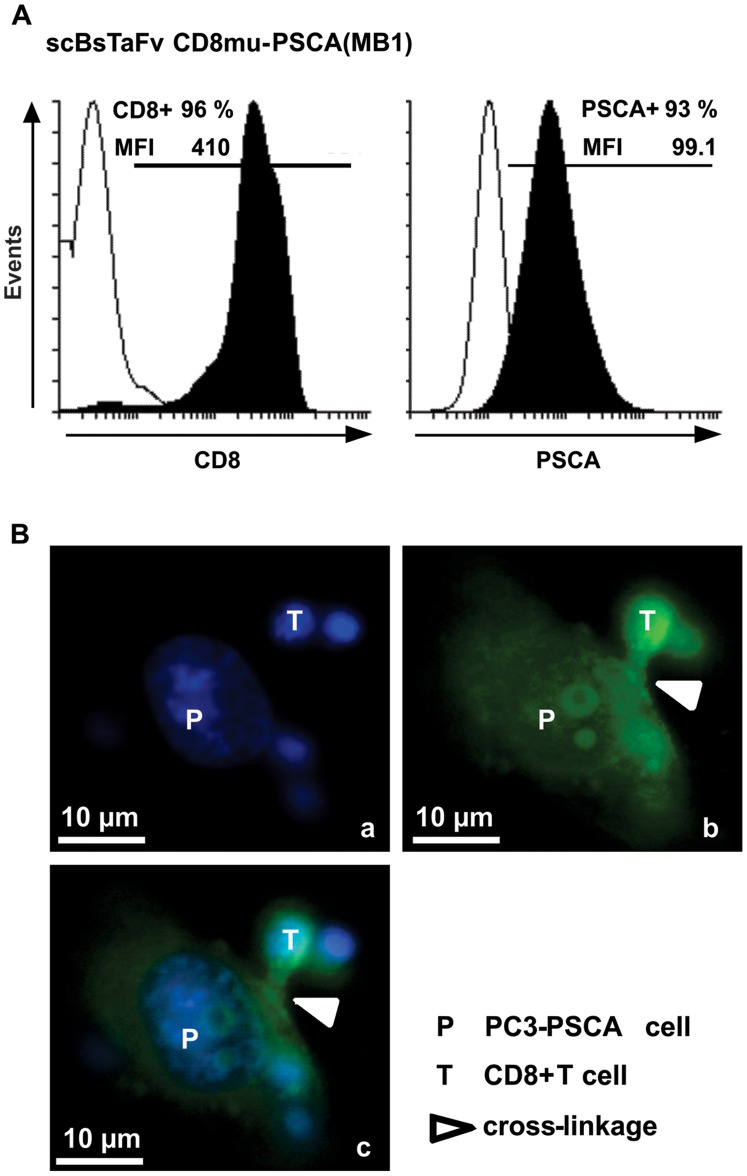
Figure 2. Binding properties of the novel scBsTaFv CD8-PSCA(MB1).
A, in order to investigate binding properties of the novel single-chain bsAb, PC3-PSCA cells and isolated CD8+ T cells were stained with 10 ng/µl of recombinant Ab. Specific binding of the scBsTaFv CD8-PSCA(MB1) was detected with anti-myc/FITC. Histograms show percentage and mean fluorescence intensity (MFI) of stained antigen-positive cells (black) in comparison to the negative control incubated only with the secondary anti-myc/FITC mAb (white). B, to demonstrate simultaneous binding of the novel scBsTaFv CD8-PSCA(MB1) to PC3-PSCA cells and CD8+ T cells and, hence, to visualize a cross-linkage between the two cell types, microscopic images were taken. Therefore, PC3-PSCA cells and isolated CD8+ T cells were co-cultivated in the presence of scBsTaFv CD8-PSCA(MB1) for 22 h and fixed with 90% methanol. The scBsTaFv CD8-PSCA(MB1) was detected with anti-myc/FITC mAb (green) and cell nuclei were stained with DAPI (blue) containing sample cover medium. Microscopic image (a) shows DAPI-stained nuclei of one PC3-PSCA cell (P) surrounded by four T cells (T). A homogenous cell surface staining of the PC3-PSCA cell and T cells is shown in picture (b) after detection of the scBsTaFv CD8-PSCA(MB1) with anti-myc/FITC. Furthermore, a cross-linkage between the PC3-PSCA cell and a T cell is visible (white triangle). Image (c) is an overlay of (a) and (b).

