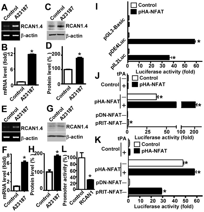
Figure 2. Calcium overloading upregulates RCAN1 isoform 4 expression through activation of NFAT signaling pathway.
(A) Calcium ionophore A23187 increased RCAN1 isoform 4 mRNA expression in HEK293 cells. A specific set of primers were used to amplify a RCAN1 isoform 4 mRNA through RT-PCR. Samples were analyzed on 1.5% agarose gel. β-actin was used as internal control. (B) Quantification of (A). Values represent means ± SEM. n = 3, *P<0. 05 by student's t-test. (C) Calcium ionophore A23187 increased RCAN1 isoform 4 expression in HEK293 cells. RCAN1.4 protein expression is upregulated by A23187. HEK293 cells were treated with 2.5 µM A23187 for 12 hours. 150 ug cell lysates were separated in a 15% glycine SDS-PAGE gel. RCAN1 was detected with anti-RCAN1 antibody DCT3. β-actin detected with anti-β-actin antibody (Sigma, AC15) served as internal control. (D) Quantification of (C). Values represent means ± SE (n = 3), *P<0. 05 by student's t-test. (E) Calcium ionophore A23187 increased RCAN1 isoform 4 mRNA expression in SH-SY5Y cells. SH-SY5Y cells were treated with 2.5 µM A23187 for 12 hours. A specific set of primers were used to amplify a RCAN1 isoform 4 mRNA by RT-PCR. The samples were analyzed with 1.5% agarose gel. β-actin was used as an internal control. (F) Quantification of (E). Values represent means ± SE (n = 3), *P<0. 05 by student's t-test. (G) Calcium ionophore A23187 increased RCAN1.4 expression in SH-SY5Y cells. RCAN1.4 protein expression was upregulated by the treatment with A23187. SH-SY5Y cells were treated with 2.5 µM A23187 for 12 hours and 150 ug cell lysates were separated in a 15% glycine SDS-PAGE gel. RCAN1.4 was detected with anti-RCAN1 antibody DCT3. β-actin served as an internal control. (H) Quantification of (G). Values represent means ± SE (n = 3), *P<0. 05 by student's t-test. (I) NFAT significantly increased pDE4luc promoter activity. NFAT expression plasmid pHA-NFAT was co-transfected with RCAN1.4 promoter pDE4luc into HEK293 cells. pGL3-Basic was used as a negative control. And pIL2Luc that contains NFAT responsive elements was used as a positive control. Luciferase activity was measured 48 hours after transfection by a luminometer. Values represent means ± SE (n = 4), *P<0. 05 by student's t-test. (J) and (K) RCAN1 exon 4 promoter is activated by NFAT independent of AP1. HEK293 cells co-transfected with pHA-NFAT and pIL2Luc (J) or pDE4luc (K) were exposed to 100 nM tPA for 20 hours. NFAT dominant negative plasmid pDN-NFAT and mutant NFAT plasmid pRIT-NFAT that cannot interact with AP1 were also co-transfected. Luciferase activity was measured with a luminometer 48 hours after transfection. The X axis indicates fold increase of luciferase activity. Values represent means ± SE (n = 4), *P<0. 05 by student's t-test. (L) RCAN1 overexpression decreased RCAN1 exon 4 promoter activity. HEK293 cells were co-transfected with pRCAN1mychis and RCAN1 promoter constructs pDE4Luc. Luciferase activity was measured 48 hours after transfection. Values represent means ± SE (n = 4), *P<0. 05 by student's t-test.

