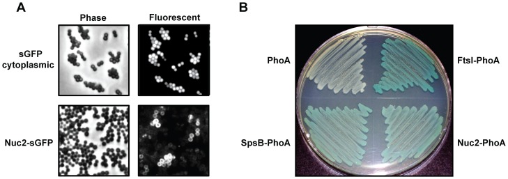
Figure 2. Localization and orientation of Nuc2 in the cell membrane.
A. Phase and fluorescent microscopy of S. aureus expressing cytoplasmic sGFP or Nuc2-sGFP translational fusion. B. Alkaline phosphatase fusions grown in a PhoA dye indicator plate assay, where growth appearing white indicates PhoA is facing the cytoplasm and blue indicates PhoA is exposed to the periplasm. Shown clockwise from the upper left corner are: the E. coli phoA mutant host, an E. coli positive control protein fusion (FtsI-PhoA), the S. aureus Nuc2-PhoA protein fusion, and a S. aureus positive control protein fusion (SpsB-PhoA).

