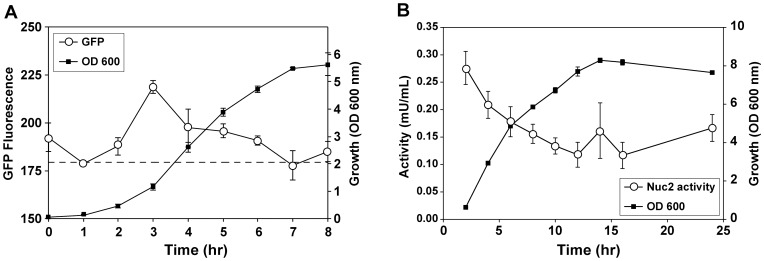
Figure 5. Nuc2 is expressed at low levels during growth.
A. Monitoring changes in fluorescence of a Pnuc2-sGFP transcriptional reporter over time (open circles) in comparison to growth (black squares). Dotted line indicates level of background fluorescence of growth media. B. Changes in surface Nuc2 activity (open circles) present in a S. aureus USA300 LAC nuc mutant over time as measured by whole-cell FRET assay.

