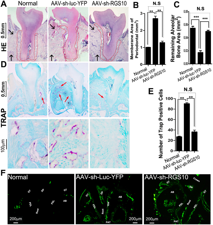
Figure 3.
AAV-mediated Rgs10 knockdown decreased bone resorption and the number of TRAP-positive osteoclasts in the periodontal area. (A) Representative figures of H&E staining from uninfected mice (Normal), W50-infected mice treated with AAV-sh-Rgs10 or AAV-sh-luc-YFP. Black arrows indicate significant change of alveolar bone in different groups, which demonstrate that the alveolar bone resorption was severe in AAV-sh-luc-YFP treatment group. And the periodontal ligament area in AAV-sh-luc-YFP treated disease group showed irregular shape compared to the normal and AAV-sh-Rgs10 treated group (red dot area). (B, C) The area of periodontal ligament increased in the AAV-sh-luc-YFP treatment group, compared to the normal and AAV-sh-Rgs10 treatment groups (P<0.01) and remaining alveolar bone area was also decreased significantly in AAV-sh-luc-YFP treated disease group (P<0.001) (N=9, n=3 per group, repeated 3 times). (D) Representative figures of TRAP staining (counter stained with fast green) from uninfected mice (Normal), W50-infected mice treated with AAV-sh-Rgs10 or AAV-sh-luc-YFP (Red Arrows). (E) Compared with normal control group, the number of TRAP positive osteoclasts from AAV-sh-luc-YFP groups increased significantly. The AAV-sh-Rgs10 treatment group has obviously therapeutic effect, and the number of osteoclasts were close to normal control group (P<0.01). (F) Fluorescence microscope image of sections reveal YFP expression after local injection with AAV-sh-luc-YFP and eGFP expression of AAV-sh-Rgs10 compared with normal mouse tissues that did not receive AAV treatment (GT: Gingival Tissue, AB: Alveolar Bone, DP: Dental Pulp, PDL: Periodontal Ligament, PAT: Periapical tissue) (N=9, n=3 per group, repeated 3 times). N.S: no significant; **P<0.01; ***P<0.001.

