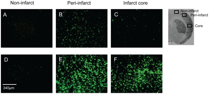
Figure 5. Microglial activation.
Immunohistochemical sections from the cortex of the ipsilateral hemisphere from an animal receiving a pre-treatment of vehicle (A–C) or Compound 21 (50 ng/kg/min) depicting microglial activation using OX42 staining (green) in the non-infarcted tissue (A and D); the peri-infarcted tissue (B and E) or the core region of damage (C and F).

