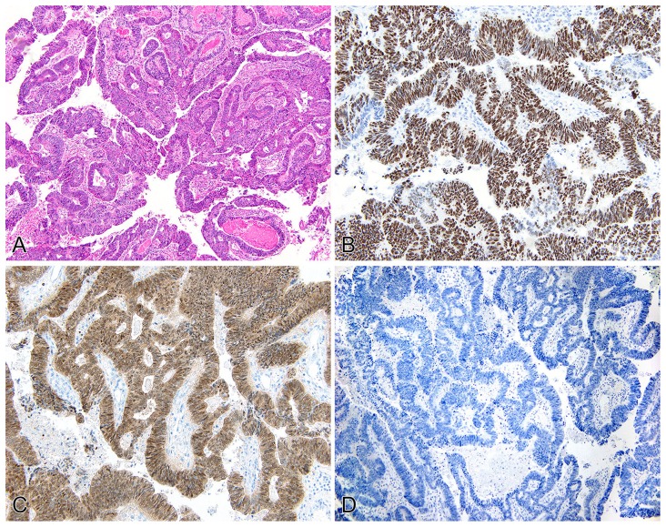
Figure 1. Primary adenocarcinoma of the urinary bladder (pT2).
H&E displaying intestinal-type architecture (A). p53 staining shows diffuse, strong nuclear reactivity (B). p16 shows both strong nuclear and cytoplasmic staining in tumor cells (C). Immunostaining for HPV shows no reactivity in any of the tumor cells (D).

