
Figure 2.
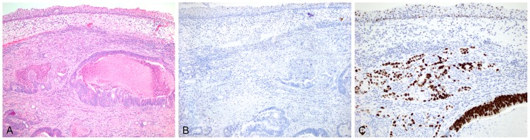
An H&E section of a separate case (pT2) shows adenocarcinoma underlying residual urothelium (A). This case shows no reactivity to p16 within the tumor or the normal urothelium with some faint nonspecific background staining seen (B). Very strong nuclear reactivity to p53 is seen in essentially all tumor cells. Weak-to-moderate reactivity is seen within the overlying urothelial cells (C).