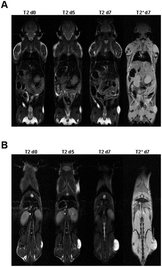
Figure 4. Development of tumor volume with and without anti-YKL-40 treatment.
T2 and T2* weighted MRI of two Balb/c scid mice with LOX tumors treated with isotype (A) or anti-YKL-40 monoclonal antibody (B) before injection of antibody (d0), five days (d5) and seven days (d7) after start of treatment. Observed hemorrhage is indicated by arrow.

