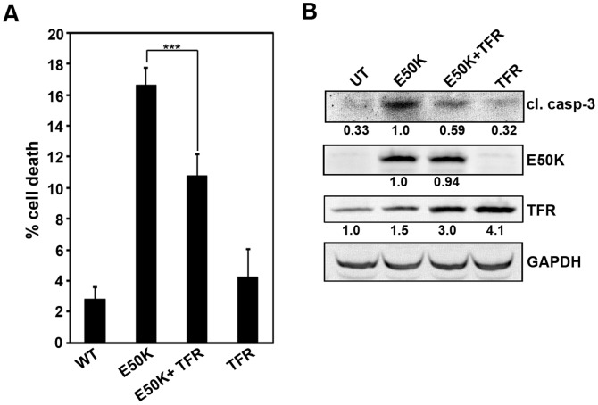
Figure 2. Effect of TFR expression on E50K-OPTN-induced cell death.
(A) The cells plated on coverslips were transfected with 100ng of GFP-E50K alone or 200ng of TFR alone or cotransfected with 100ng GFP-E50K and 200ng TFR. After 32 hours of overexpression, cells were stained with TFR antibody and cell death was analysed. Data shown here represent cell death in expressing cells after subtracting background cell death from non-expressing cells from 6 experiments (mean±SD). ***p<0.001 (student’s t-test) (B) Cells plated in 35-mm dishes were transfected with 1 µg of GFP-E50K alone or 2 µg of TFR alone or cotransfected with 1 µg GFP-E50K and 2 µg TFR. After 32 hours of overexpression, cell lysates were made and subjected to western blotting with cleaved caspase-3, GFP and TFR antibodies. GAPDH was used as a loading control. UT, untransfected.

