
Figure 4. Involvement of autophagy in E50K-induced cell death.
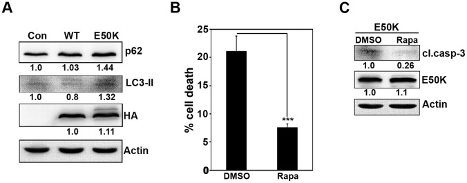
(A) Cells plated in 24-well plate were infected with control or WT OPTN or E50K adenoviruses and allowed to express for 24 hours. Protein lysates were subjected to western blotting with p62, LC3-II and HA antibodies. Actin was used as a loading control. (B) Cells plated on coverslips were transfected with GFP-E50K plasmids. After 12 hours of transfection, cells were treated either with DMSO or with 1 µM rapamycin (Rapa) for 20 hours. At the end of 32 hours of overexpression, cells were fixed and mounted to score for cell death. Bar diagram depicts percentage of cells showing E50K-induced cell death in the presence and absence of rapamycin. Data represents mean ± SD of 5 experiments. ***p<0.001 (Student’s t-test). (C) Cells plated in 35-mm dishes were transfected with GFP-E50K and after 12 hours of transfection, cells were treated with either DMSO or rapamycin (Rapa) (1 µM) for 20 hours. At the end of 32 hours of overexpression, lysates were made and subjected to western blot with cleaved caspase-3 and GFP antibodies. Actin was used as a loading control.