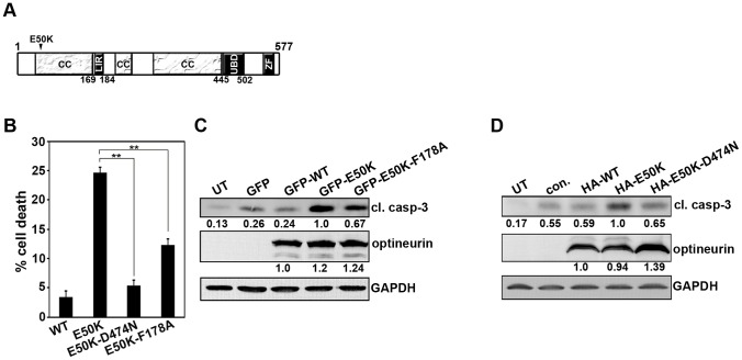
Figure 5. E50K-OPTN-induced cell death is dependent on UBD and LC3-interacting region.
(A) Schematic of optineurin protein showing domains LIR (LC3 interacting region) and UBD (ubiquitin binding domain), ZF (zinc finger) and CC (coiled coil). (B) Cells plated on coverslips were transfected with 300ng of GFP-E50K or GFP-E50K-D474N or GFP-E50K-F178A. After 32 hours of transfection, cells were fixed and cell death was determined in the respective expressing cells. Bar diagram showing quantitation of cell death in the presence of each of the mutants after subtracting background cell death in non-expressing cells. Data represent mean ± SD of 3 experiments. **p<0.01 (Student’s t-test) (C) Cells were transfected with 3 µg of GFP control plasmid or GFP-WT or GFP-E50K or GFP-E50K-F178A plasmids and overexpressed for 32 hours. Western blot was done with cleaved caspase-3 and GFP antibodies. GAPDH was used as a loading control. UT, untransfected (D) The cells were transfected with 3 µg of control plasmid or HA-WT or HA-E50K or HA-E50K-D474N plasmids and overexpressed for 32 hours. Western blot was done with cleaved caspase-3 and HA antibodies.

