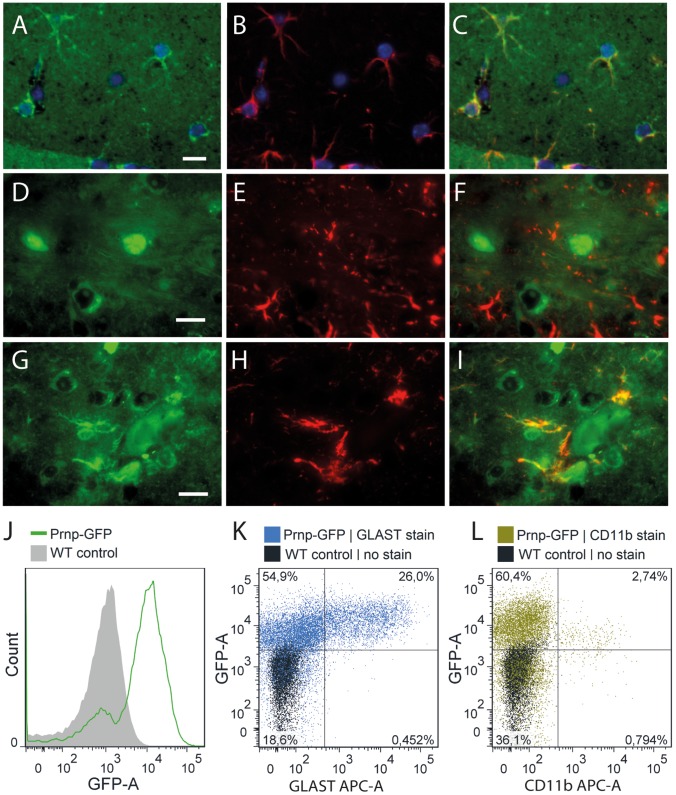
Figure 4. Immunolocalization of PrP, GFP and GFAP in ki-Prnp-GFP +/k brain.
(A) In uninfected brains, GFP is observed in astrocyte-shaped cells, which are GFAP+ (B, C). In order to increase the astrocyte size and therefore enhance staining and imaging, brains with middle stage (week 41 of 54 incubation course) prion-induced reactive gliosis were studied (D–I). (D) PrP (green) is mostly diffusely extracellular and intracellular in some neurons, but does not co-localize with GFAP (red) of astrocytes (E, F). Nuclei were counterstained with DAPI. (G) GFP is present in some cell bodies and co-localizes with GFAP (E, F). (J–L) FACS analysis of dissociated ki-Prnp-GFP brains. (J) Histogram distribution of GFP fluorescence in ki-Prnp-GFP k/k mice shows distinct GFP- and GFP+ populations, comprising ∼17% and ∼83% of cells respectively. (K) Most GLAST+ cells are GFP+ and their GFP fluorescence intensity is higher than for GLAST- cells (medians of populations are 0.6×10⁴ for the top left quadrant, and 1.5×10⁴ for the top right quadrant). (L) CD11b+ cells show lower GFP fluorescence intensity then CD11b− cells (medians of populations were 1.1×10⁴ for the top left quadrant, and 0.57×10⁴ for the top right quadrant). Scales in A, D and G represent 10 µM.

