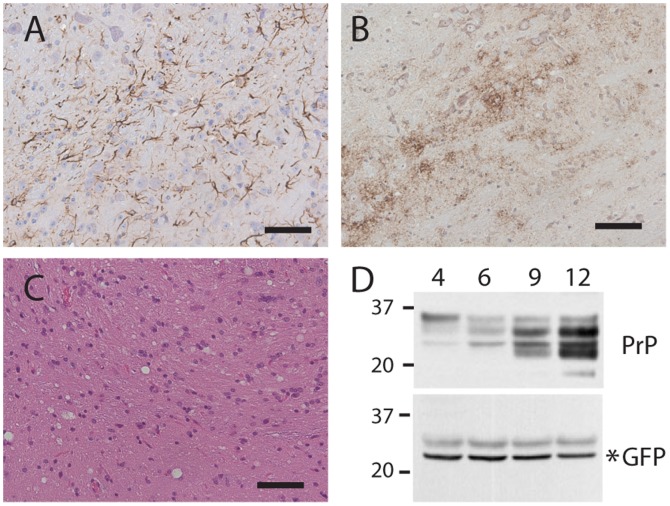
Figure 5. RML scrapie infection induces accumulation of PrP but not GFP in heterozygous ki-Prnp-GFP/WT mice.
Reactive gliosis (A), PrPres (B), and spongiosis (C) are apparent in brains following a 41 week (9 months) incubation. Brains harvested at 4, 6, 9, and 12 months after infection show increasing PrP (D, top blot), but not GFP (D, lower blot). Molecular weight markers are on the left, and time post infection (months) is indicated above the blots. Scale bars in A–C represent 50 µM.

