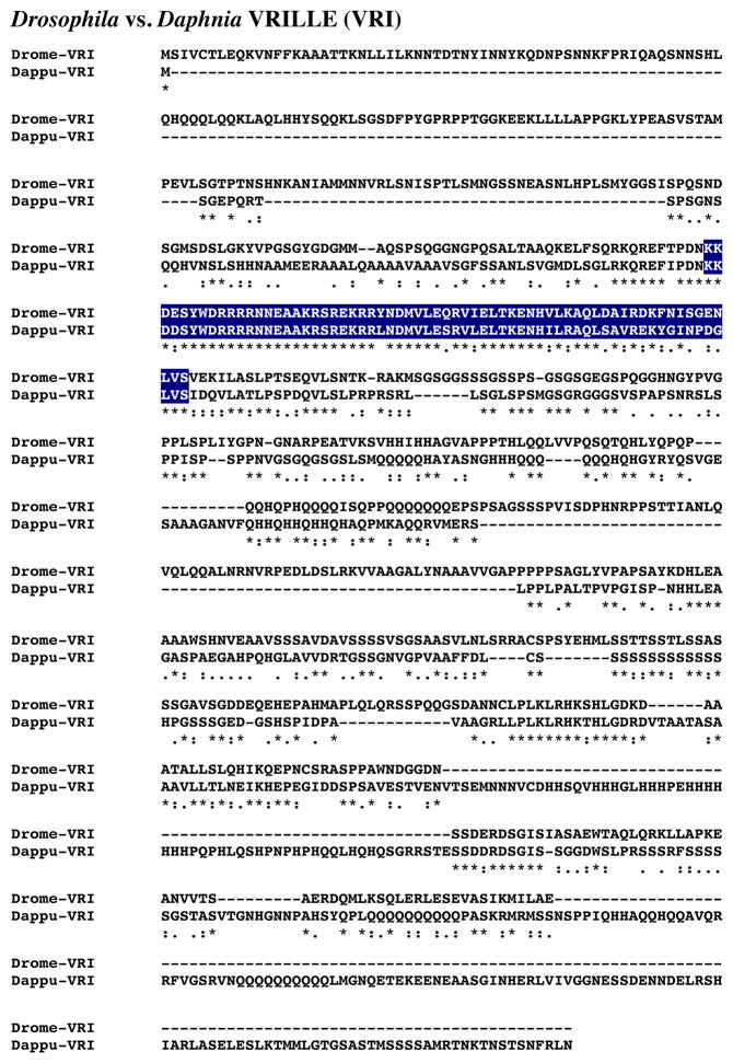
Figure 13.
Putative Daphnia pulex VRILLE (VRI) protein. Alignment of Drosophila melanogaster VRI (Drome-VRI) with D. pulex VRI (Dappu-VRI). In the line immediately below each sequence grouping, stars indicated amino acids that are identically conserved, while single and double dots denote amino acids that are similar in structure. In this figure, basic region leucine zipper domains identified by SMART analyses are highlighted in dark blue.

