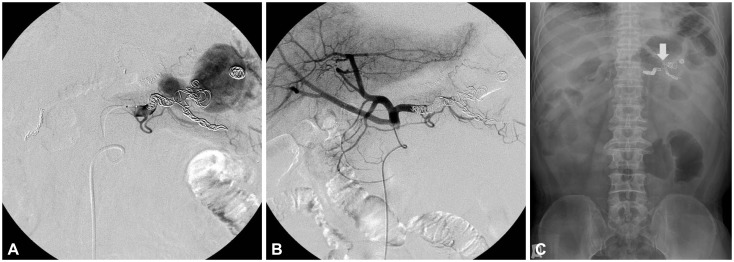
Fig. 2.
(A) Embolization of the pseudoaneurysm was done by using coils. (B) Angiography after coil embolization revealed total occlusion of the pseudoaneurysm. (C) Simple abdominal radiograph taken just after embolization showed the multiple coils (arrow) placed within the pseudoaneurysm and splenic artery.

