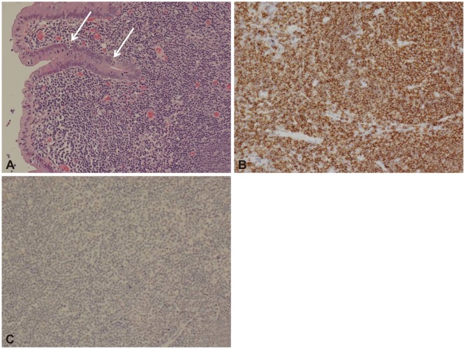
Fig. 2.
Histologic findings. (A) Lymphoma cells, with small to medium-sized nucleoli with pale cytoplasm, infiltrated the reactive follicles. The epithelium was invaded and destroyed by discrete aggregates of lymphoma cells (arrows) (H&E stain, ×200). (B) Immunohistochemical staining revealed lymphoma cells with a diffuse immunoreaction for CD20 (Immunostain for CD20, ×200). (C) Immunohistochemical staining revealed that lymphoma cells were not immunoreactive for cyclin D1 (Immunostain for cyclin D1, ×200).

