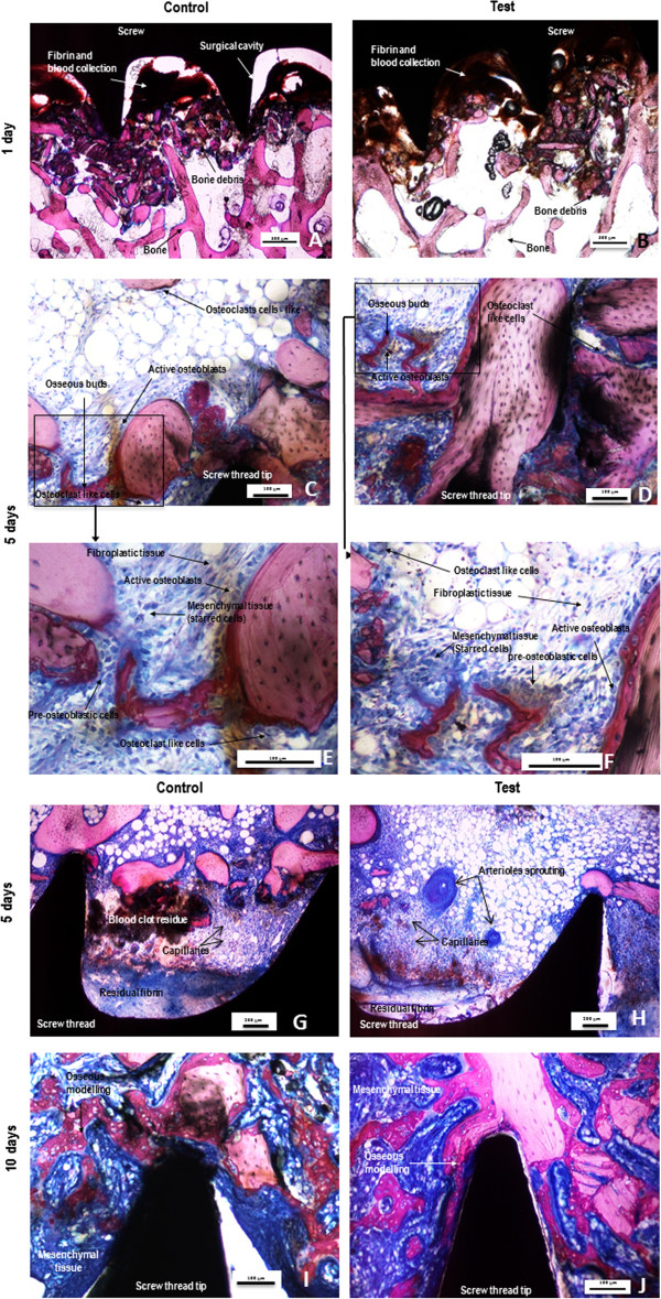
Figure 3.

Representative photomicrographs of undecalcified sections of control (A, C ,E, G and I) or test (ZOL coated) (B, D, F, H and J) screws after 1 (A and B), 5 (C,E,G and D,F,H) and 10 (I and J) days stained with a modified paragon. Photomicrographs E and F are respectively high power views of photomicrographs C and D (frames). The signs of repair with formation of fibro-mesenchymal tissue and woven bone are increased in the test groups without evidence of cellular adverse effects. Yet at 5 days, the test group (picture H) showed more advanced signs of angiogenesis than the control group (picture G). Osteoclasts remained active around the test implants.
