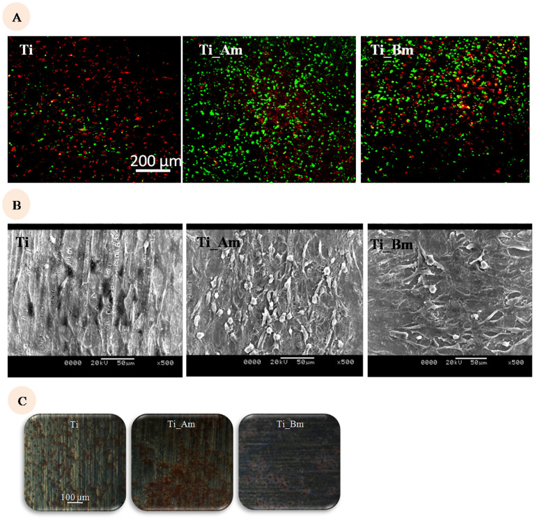
Figure 6. Effect of macrophage-osteoblast coculture on osteoblast adhesion and function.
(6A)-Influence of macrophage, on osteoblast adhesion on Ti surface. Pre-stained macrophage (red) and osteoblast (green) are seeded on pristine and modified Ti surface in 2:1 ratio. Imaging with CLSM is done after 24 hr. (6B)-Influence of macrophage, on Osteoblast morphology. Macrophage and osteoblast are seeded on pristine and modified Ti surface and subjected to scanning electron microscopy. Scale bar is 50 μm. (6C)-Calcium deposition on the co-cultured surfaces. Imaging of alizarin red stained co-cultured surfaces is done after 14 days. Scale bar is 100 μm.

