Figure 5.
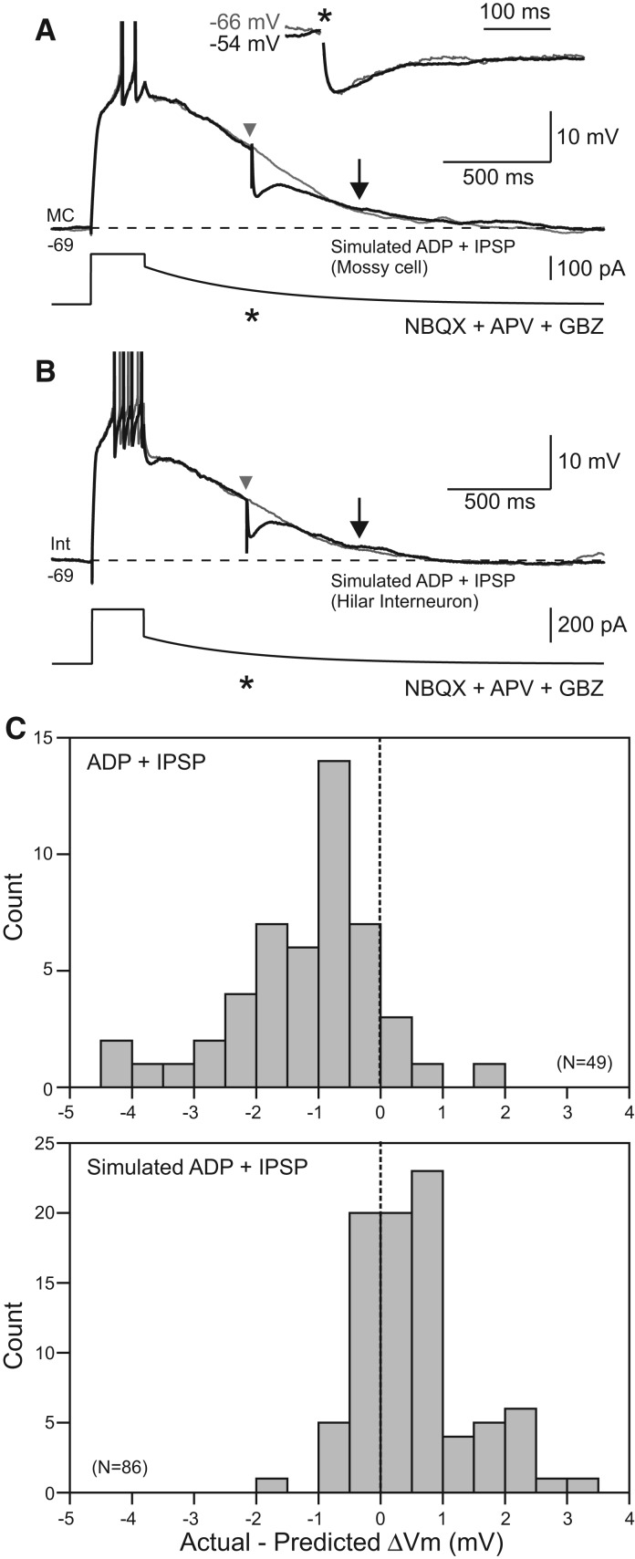
IPSPs do not affect simulated afterdepolarizations. (A) Mossy cell responses to slowly decaying current injection designed to mimic afterdepolarization responses in control conditions. IPSPs evoked during simulated ADPs (black trace, timing indicated by an asterisk [*]) did not decrement the membrane potential beyond the drop predicted by combining the simulated ADP and IPSP responses evoked in isolation. Arrows indicate response time points analyzed. (Inset) Similar kinetics of evoked IPSPs at two different membrane potentials in a different mossy cell. IPSPs scaled to the same peak amplitude. (B) Similar protocol evoked in a hilar interneuron also failed to show an effect of evoked IPSPs on simulated ADPs. APs truncated in A and B. (C) Histograms of modulation of real (top) and simulated (bottom) afterdepolarization responses by evoked IPSPs. Values indicated reflect modulation of membrane potential beyond the voltage predicted by analyzing the ADP and IPSP responses evoked in isolation. The two distributions were statistically different (P < 0.0001, two-sample Kolmogorov–Smirnov test).

