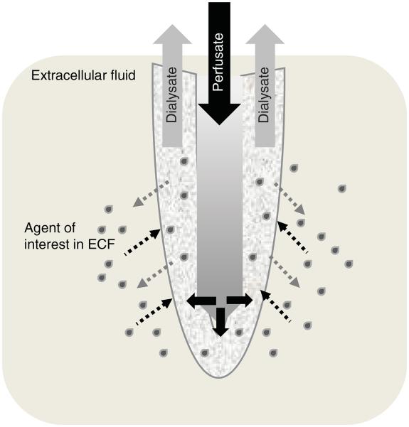
Figure 2. Schematic of microdialysis catheter membrane.
Schematic depiction of the probe portion of a microdialysis catheter (not drawn to scale). The semi-permeable membrane at the tip of the probe allows diffusion of fluid (perfusate out and ECF in) along concentration gradients. Perfusate enters the central portion of the probe under a constant flow rate (i.e., 1 μl/min). It then moves to the outer compartment of the probe where it is admixed with the ECF and the solute under investigation (grey drops) is collected in the outlet tubing.
ECF: Extracellular fluid.

