
Figure 1.
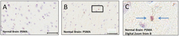
PSMA staining in tissues samples representative of normal brain. A – Normal brain showing no PSMA staining. B – Normal brain probed for PSMA showing no blood vessel staining and light staining of cellular elements, magnified digitally in (C) and indicated by arrows.