
Figure 5.
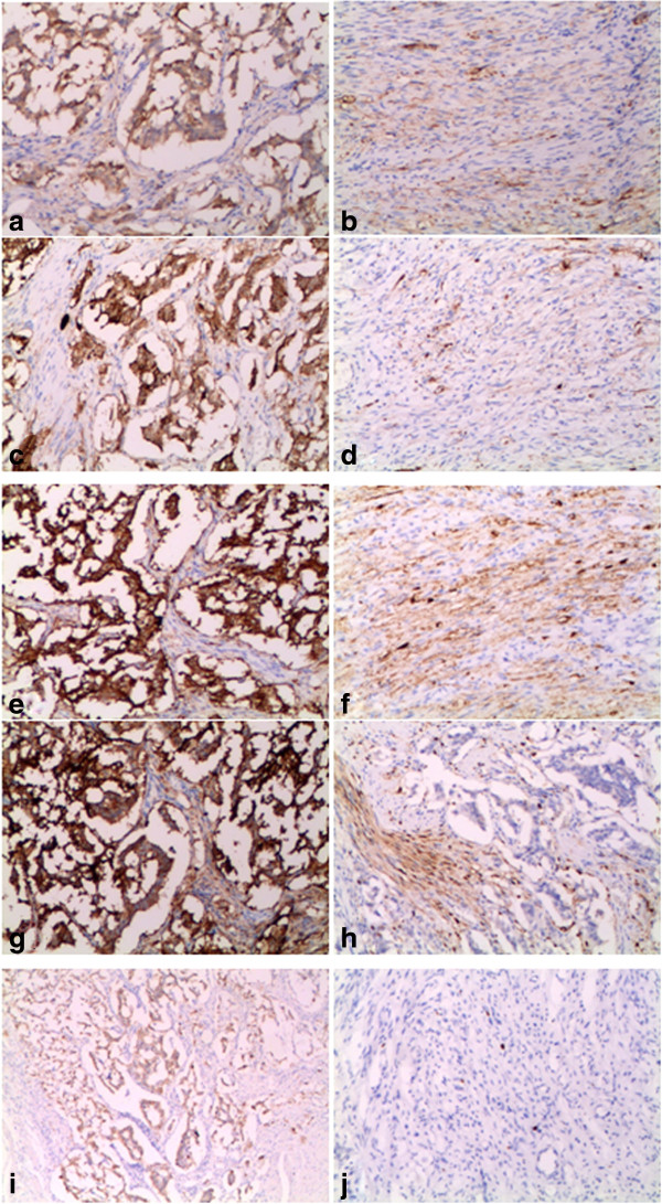
Immunohistochemistry staining of the tumor tissue. Epithelioid cell nests are positive for NSE (a), CgA (c), Syn (e), CD56 (g), CK (i). Spindle cells are positive for NSE (b), CgA (d), Syn (f), S-100 (h). Ki-67 staining shows the proliferative rate is less than 1% (j).