Figure 2.
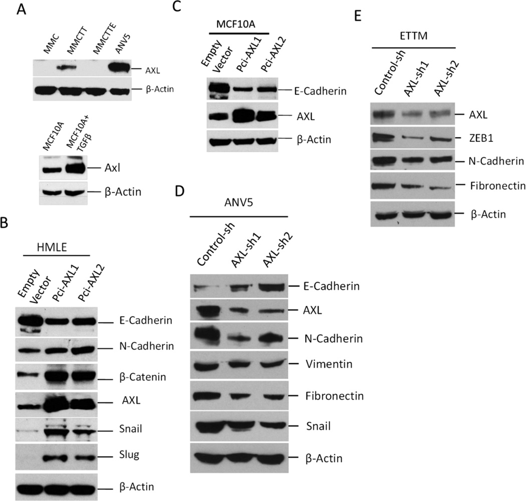
AXL overexpression regulates expression of EMT markers. A, Immunoblot analysis showing the effect of TGFβ/TNFα-mediated EMT induction on AXL expression in MMC. MMCTT are MMCs treated with TGFβ and TNFα MMCTTE are MMCTT mesenchymal cells that reverted back to epithelial cells following treatment withdrawal. ANV5 are shown as control. Also shown is AXL expression in MCF-10A following treatment with TGF-β. B–C, Immunoblot analyses of EMT marker expression in HMLE and MCF10A. Overexpression of AXL was induced in HMLE and MCF10A using two separate expression vectors, pCi-AXL1 and pCi-AXL2. D–E, Silencing of AXL in ANV5 and ETTM cells reverses mesenchymal phenotype of ANV5 cells characterized by increased E-cadherin expression but decreased expression of AXL, N-cadherin, Vimentin, Fibronectin, Zeb1 and Snail. Control cells expressed a scrambled shRNA. The results of the experiments in this figure were repeated independently two times with similar results.

