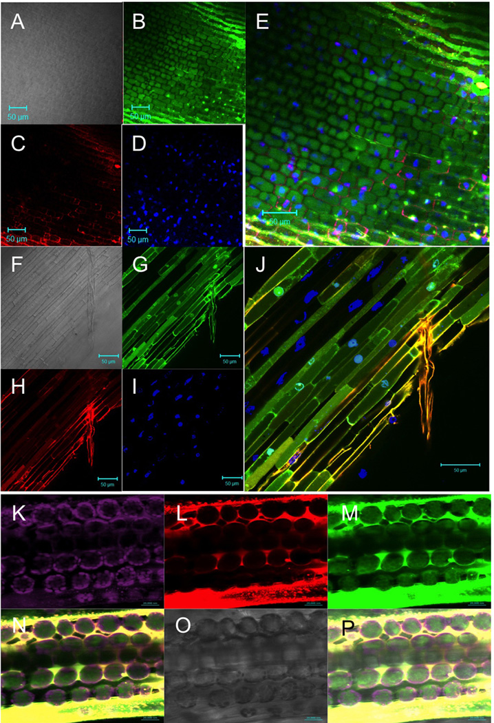
Fig. 2.
Specific microbe-independent entry of Avr1bNt proteins into soybean root cells and wheat leaf cells. A to E, Soybean root entry by Avr1b N-terminus (residues 1–50) labeled by Dylight488, and counter stained with propidium iodide and 4',6-diamidino-2-phenylindole (DAPI). F to J, Soybean root entry by Avr1b N-terminus (residues 1–50) labeled by Dylight488, mixed with Avr1b N-terminus with RFLR->qFLR mutation labeled with DyLight550, and counter stained with DAPI. A,F, light image; B,G, Dylight488 image; C, propidium image; H, Dylight550 image; D and I, DAPI image; E and J, overlay of the three fluorescent images. In A to J, purified fusion proteins (0.4 mg/ml each) were incubated with soybean (Williams) root tips, for 2 hours at 25°C in PBS buffer adjusted to pH 7.2, then washed for 15 min in formalin (PBS + 10% formaldehyde) containing 0.2 µg/ml propidium iodide (A to E only) and 0.4 µg/ml DAPI. K to P, Wheat leaf cell entry by Avr1b N-terminus (residues 1–50) labeled by Dylight488 mixed with Avr1b N-terminus (residues 1–50) with RFLR->qFLR mutation labeled by Dylight550. Purified fusion proteins (0.4 mg/ml each) in PBS buffer adjusted to pH 7.2 were infiltrated with a blunt syringe into ~9 day old wheat seedling leaves. Leaves were imaged after 6 h without washing. K, chloroplast fluorescence (excitation 488 nm; emission meta filter 675–715 nm); L, Dylight550 image (excitation 543 nm; emission window 585–615 nm; gain 600 to 654; digital offset −0.1 to 0); M, Dylight488 image (excitation 488 nm; emission window 505–530 nm; gain 580 to 610; digital offset −0.1 to 0); N, overlay of K to M; O, light image; overlay of N and O. Labeling of proteins with Dylight dyes was as described (Sun et al., 2013). The experiments shown in this figure were conducted at Virginia Tech using a Zeiss LSM 510 Meta confocal microscope.

