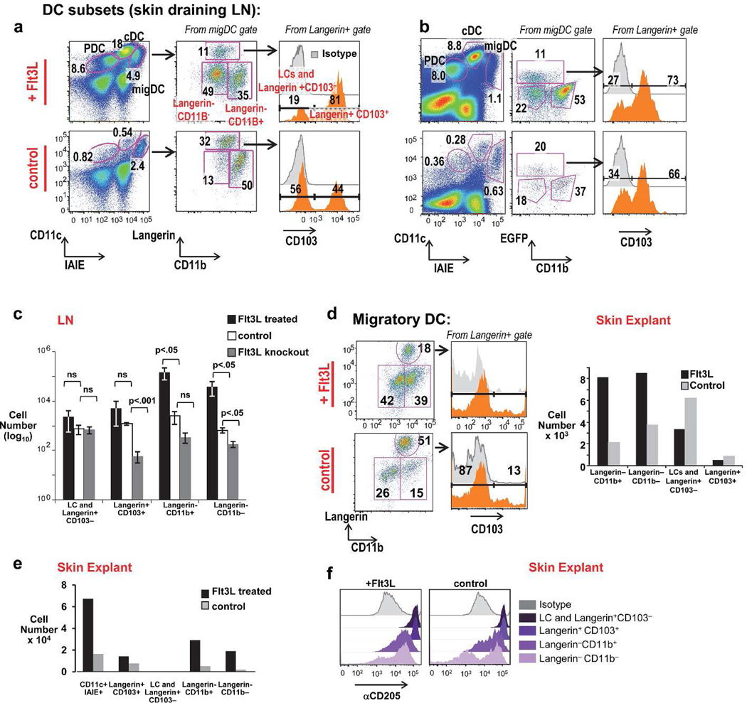
Figure 1. Flt3L dependence of migratory DC subsets, including Langerin-CD11b− DC.
A) Schema of LN DC from Flt3L-tumor treated vs. C57Bl/6 (B6) control mice at day 13. PDC (plasmacytoid DC), cDC (classical LN resident DC), migDC (migratory DC) are labeled. (B) LN DC subsets Flt3L-treated (upper) vs. untreated Langerin-GFP mice. (C) Quantitation of migratory DC from Flt3L-treated, control, Flt3L−/− mice (quantitated per skin draining LN; error bars show mean +/− SD of 3 individual mice per group, analyzed by an unpaired t-test). (D) Flt3L-induced expansion of skin resident DC subsets from "crawl-out” skin explants of doxycycline-inducible Flt3L mice given 8 days of doxycycline in their drinking water (top) vs. untreated controls (bottom) (one representative experiment of two). (E) Quantitation of DC isolated from skin explants “crawlouts” of Flt3L-treated vs. control Langerin GFP reporter mice (pooled from the ears of 3 mice per experiment, one representative experiment of three). (F) α CD205+ staining of skin DC subsets from explant cultures of doxycycline treated (Flt3L+) vs. untreated (control) mice (pooled from n=2 mice per condition).

