Figure 5.
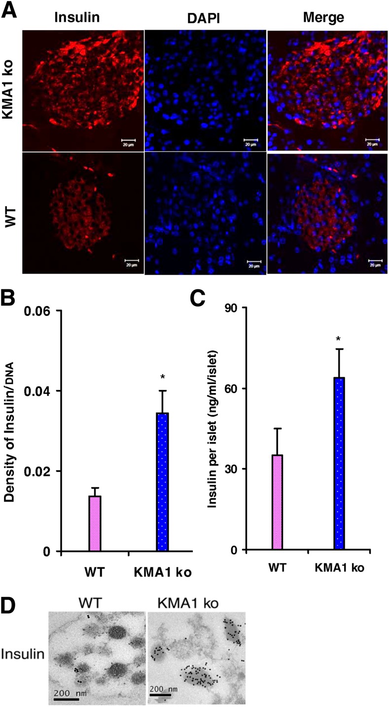
Insulin content in pancreatic β-cells. Mice were injected with 40 mg/kg/day tamoxifen in 100 μL peanut oil for 5 consecutive days, and the animals were killed 28 to 31 days later. A: Pancreatic sections were stained with anti-insulin antibody and further stained using a TRITC-conjugated anti-guinea pig antibody; DAPI was used to stain the nucleus. B: Average intensity of insulin staining relative to nuclear DAPI staining. C: Insulin content in the freshly isolated islets from KMA1ko and WT mice. D: Electron micrograph shows insulin-protein A–gold staining of insulin granules within the selected area of β-cells. *P < 0.05 (n = 3 to 4 mice per group).

