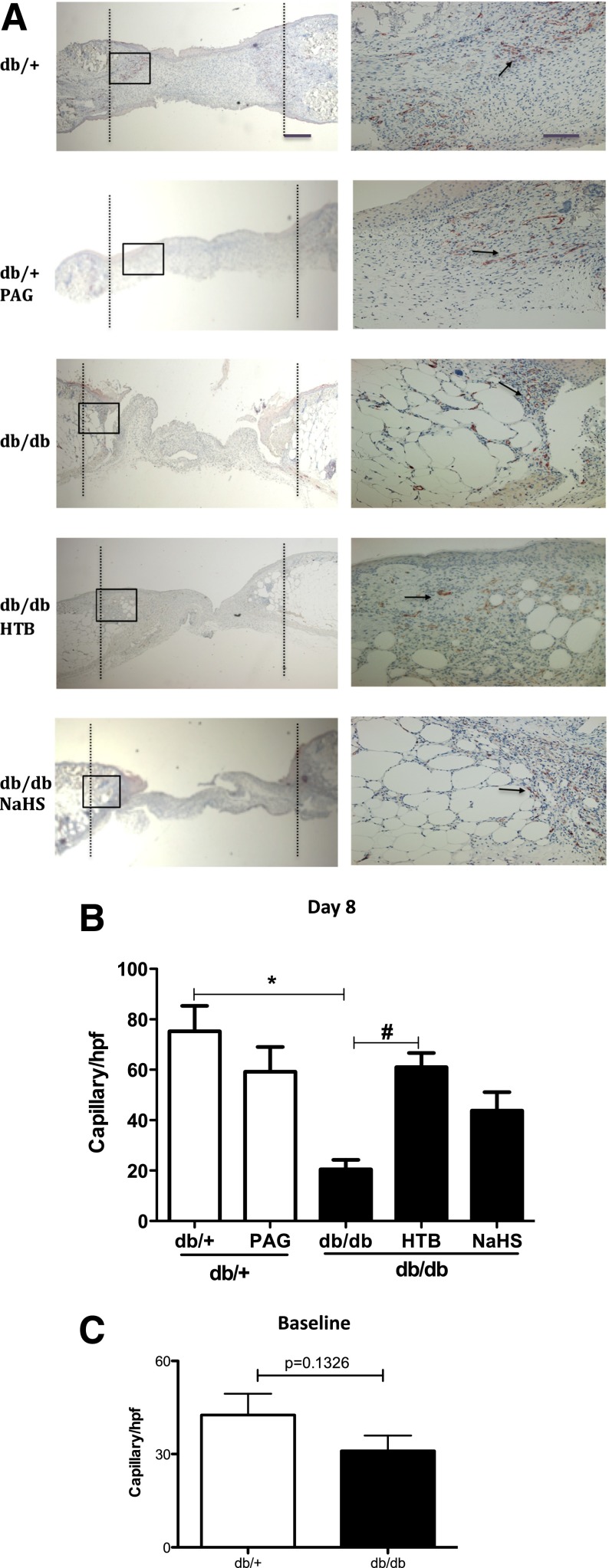
Figure 3.
H2S donor stimulated angiogenesis in wound skin from db/db mice. The granule skin tissue within the wound area was collected at day 8 after NaHS, HTB, or PAG treatment and stained with CD31. A: Representative photomicrographs of new capillaries in five groups at original magnifications ×4 (left) and ×20 (right). The black dashed lines indicate the original wound edges, the black boxes indicate the areas enlarged correspondingly in the ×20 magnification panels, and the black arrows point to CD31-positive capillaries. B: Comparison of capillary numbers per high-power field (hpf) in five groups (n = 5 in each group). *P < 0.05 vs. db/+ control, #P < 0.05 vs. db/db control. C: Comparison of capillary number in unwounded skin in db/+ mice and db/db mice (n = 5 each group).

