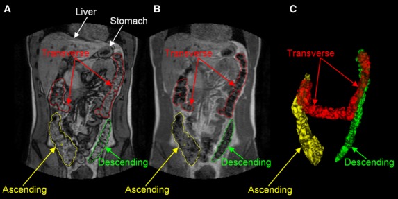
Figure 1.

Representative example of the anatomical segmentation of the colon on coronal magnetic resonance imaging (MRI) images. The left panel (A) shows the dual echo MRI image with water and fat imaged out-of-phase and the manual drawings of the regions of interest around the colon; the central panel (B) shows the corresponding MRI image with water and fat imaged in-phase; the right panel (C) shows the 3D reconstruction of the colon.
