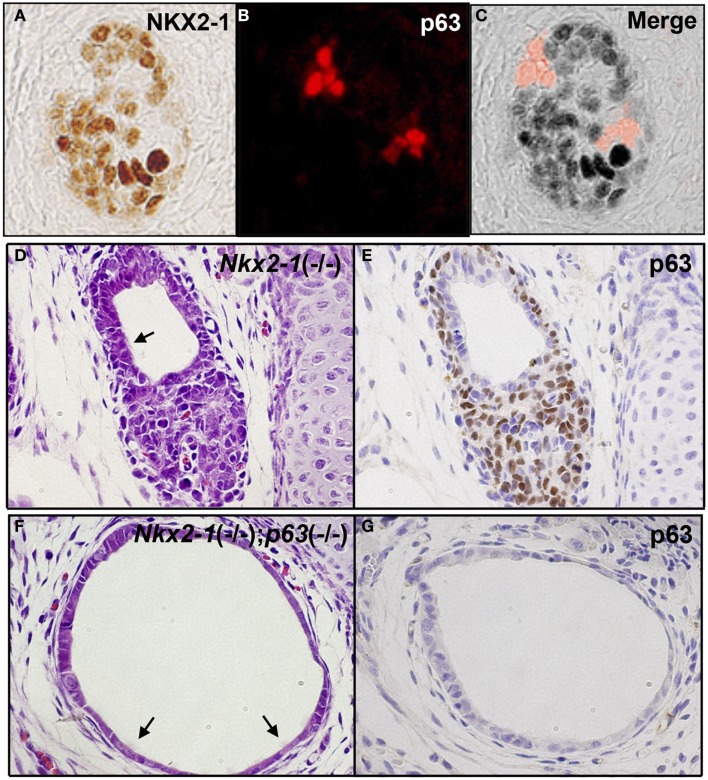
Figure 3.
p63 expression in UBB and SCN of thyroid. (A–C) Immunofluorescence of E13.5 UBB of wild-type mouse embryos for NKX2-1 (A) and p63 immunohistochemistry (B), and merged image (C). Majority of cells express NKX2-1 while a few cells express p63, both being without overlap. (D,E) SCN from E18.5 Nkx2-1-null embryos for H&E (D) and p63 immunohistochemistry (E). p63 is expressed in the stem/basal cell patterns. (F,G) SCN from E18.5 Nkx2-1; p63-double null embryos for H&E (F) and p63 immunohistochemistry (G). Note that the monolayer of p63-negative cells remain in SCN from Nkx2-1; p63-double null embryos. Arrows indicate ciliated cells observed in the cystic structure.

