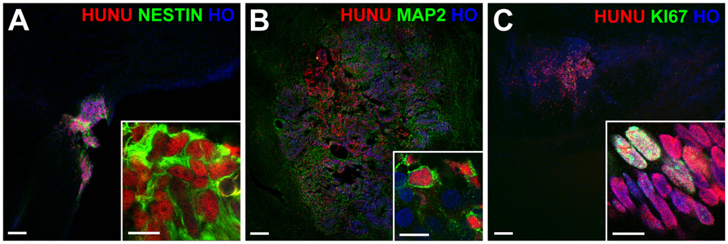
Figure 2.

Rat brain sections with live graft cells. The red-labeled human nuclei (HUNU) of graft cells also co-labeled green for the neural progenitor marker nestin (A), and, to a lesser extent, the neuronal marker MAP2 (B) and the marker of dividing cells KI67 (C). All nuclei are labeled blue with Hoechst (HO); scale bars = 100 um (10 um for the insets).
