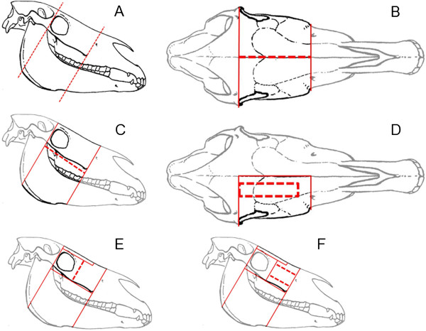
Figure 1.

Illustration of the dissection of cadaver heads. The broken red lines show the actual cut, whereas the continuous red lines show the intersections: two transversal cuts, one rostral of the Crista facialis and another caudal to the orbital cavity (A), a median sagittal cut (B), a horizontal cut parallel to the Crista facialis (C), a frontonasal bone flap (D), removal of the orbital cavity (E) and a maxillary bone flap (F) were done.
