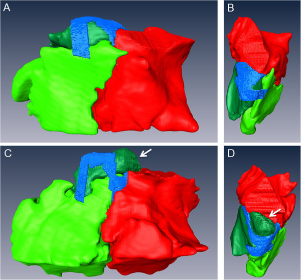
Figure 6.

3D models of the sinonasal channels and the SMR, SCV and SMC of two different horses. The sinonasal channels (blue) and the SMR (bright green), SCV (dark green) and SMC (red) of the left side of a 20-year old Warmblood gelding (A), (B) and a 20-year old Warmblood mare (C), (D) are shown in a lateral view (A), (C) and dorsal view (B), (D). Please note the large, caudo-dorsal oriented protrusion (Bulla septi sinuum maxillarium) of the SCV in the second horse (D), which leads to a subdivision of the caudal sinonasal channel (Canalis sinunasalis caudalis).
