Figure 1.

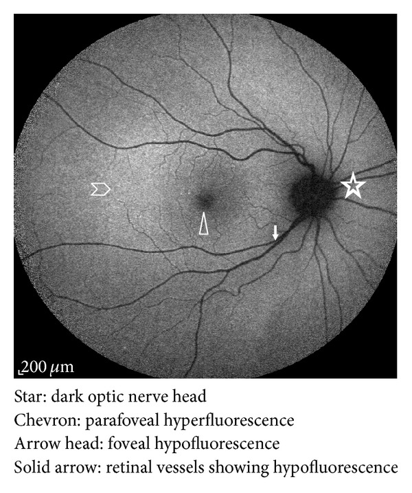
Autofluorescence distribution in a normal eye fundus. It is the highest in the posterior pole and gradually diminishes toward the periphery; it also shows hypoautofluorescence over the fovea, the optic nerve head, and retinal vessels.

Autofluorescence distribution in a normal eye fundus. It is the highest in the posterior pole and gradually diminishes toward the periphery; it also shows hypoautofluorescence over the fovea, the optic nerve head, and retinal vessels.