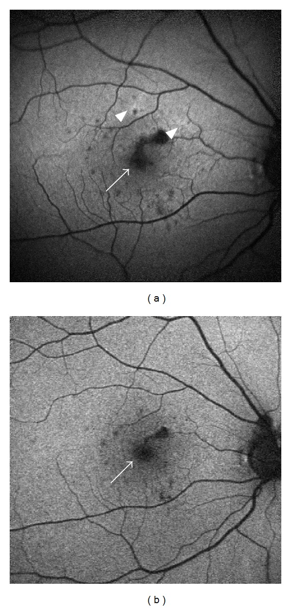
Figure 2.

Fundus autofluorescence (FAF) images of a young female with punctate inner choroiditis. (a) FAF shows hyperautofluorescence halos (arrow heads) and multiple hypoautofluorescent spots (arrow). The spots are surrounded by a hyperautofluorescent halo, denoting continued cellular damage and ongoing active inflammation. (b) FAF captured 5 months after immunosuppression was started shows diminished hyperautofluorescence and less hypoautofluorescent spots.
